Anatomy Structure Function Biology Diagrams Learn how neurons and muscles communicate through the neuromuscular junction, a type of synapse that uses acetylcholine as a neurotransmitter. Explore the anatomy, histology and clinical significance of this key component of the nervous system.
Understanding the Neuromuscular Junction. At its core, the neuromuscular junction is the connection point between a motor neuron and a muscle fiber. Imagine it as an electrical outlet in your home, where the electricity (in this case, neural signals) energizes an appliance (muscle movement).

Physiology, Neuromuscular Junction Biology Diagrams
Keywords: Neuromuscular junction, motor neuron, muscle, active zone, acetylcholine receptors, MuSK, voltage-gated calcium channels Neuromuscular junctions and motor nerves Neuromuscular junctions (NMJs) are excitatory chemical synapses formed between nerve terminals of spinal cord motor neurons and skeletal muscle fibers that use acetylcholine A neuromuscular junction, sometimes referred to as a myoneural junction, is the chemical synapse formed between a motor neuron and a muscle. Muscle Detail | Anatomy | Anatomy Of Muscle | Lower Limb Muscle. Plantaris Muscle. By Nitesh Patel - Physiotherapist January 13, 2021 August 16, 2023.
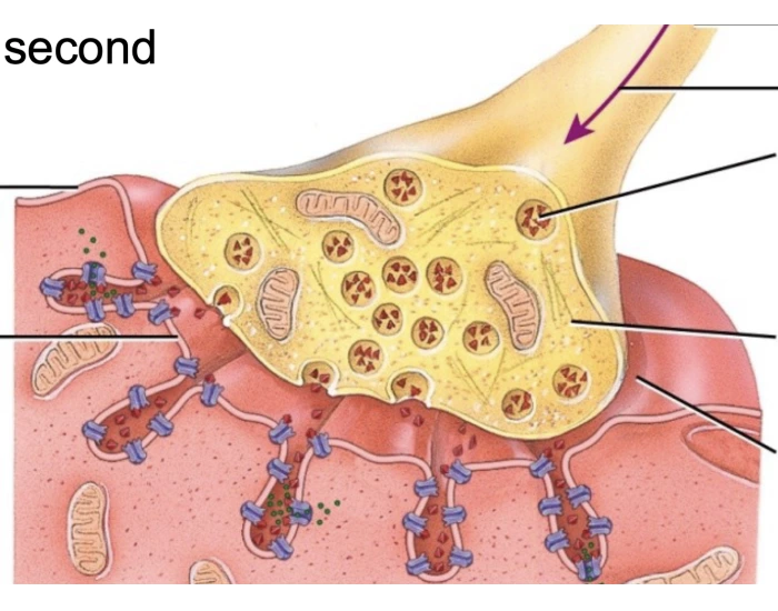
The neuromuscular junction is a synapse for the transmission of a signal from the motor nerve terminal to a postsynaptic region on the muscle fibre. It is the archetypal synapse, in the sense that it is so easy to identify and study that a lot of our understanding of synaptic neurotransmission comes from studies of neuromuscular synapses. The main molecular protagonist is acetylcholine, and it

Neuromuscular Junction Biology Diagrams
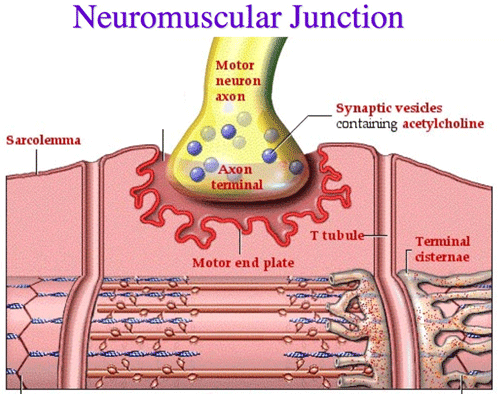
neuromuscular junction, site of chemical communication between a nerve fibre and a muscle cell.The neuromuscular junction is analogous to the synapse between two neurons.A nerve fibre divides into many terminal branches; each terminal ends on a region of muscle fibre called the end plate.Embedded in the end plate are thousands of receptors, which are long protein molecules that form channels

The neuromuscular junction (NMJ) is a synaptic connection between the terminal end of a motor nerve and a muscle (skeletal/ smooth/ cardiac). It is the site for the transmission of action potential from nerve to the muscle. It is also a site for many diseases and a site of action for many pharmacological drugs.[1][2][3][4] In this article, the NMJ of skeletal muscle will be discussed.

Practical anatomy of the neuromuscular junction in health and disease Biology Diagrams
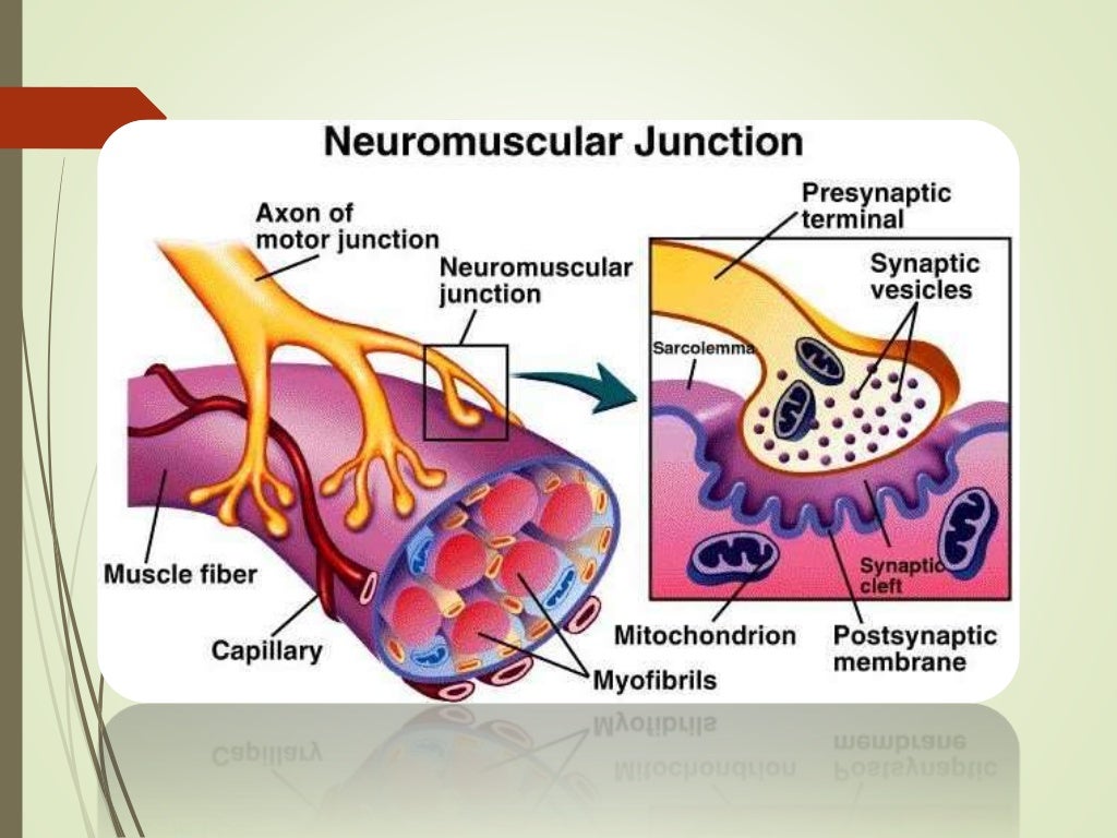
The anatomy of a neuromuscular junction can be divided into three parts: the presynaptic terminal (i.e. the motor neuron) the synaptic cleft; the postsynaptic membrane (i.e. the membrane of the muscle cell). Presynaptic Terminal. A motor neuron has a dendritic end and an axonal end. The dendrites receive the signals from adjacent neurons